
Diagnostic Imaging as the Cornerstone of Orthopedic Care
Precise diagnosis serves as the foundation for effective orthopedic recovery. Instead of relying on guesswork, medical professionals utilize diagnostic imaging to visualize structural abnormalities, such as fractures, herniated discs, or spinal stenosis, which may be the root cause of persistent neck, back, or sciatica pain. These tools provide the necessary visual evidence to distinguish between conditions that respond to conservative management and those requiring more intensive care.
The Orthopedic Spine Institute of St. Louis prioritizes a conservative-first approach, ensuring that every patient receives a tailored treatment plan based on objective clinical data. By integrating high-resolution imaging with a thorough physical examination, Dr. David S. Raskas and his team can accurately assess the severity of injuries and determine if non-surgical interventions, such as physical therapy, are sufficient. This patient-centered philosophy ensures that minimally invasive surgical options are only considered when they offer the clearest path to recovery.
Understanding your own diagnostic path starts by accessing reliable resources and professional guidance. Patients can book an appointment through our digital portal to explore their symptoms and discuss how imaging might support their journey toward improved spinal health.
Primary Imaging Tools for Spinal Assessment
 Precise diagnostics serve as the foundation for effective orthopedic recovery, and selecting the correct imaging modality is a critical step in osistl.com. Physicians typically begin with conventional X-rays to evaluate bony structures, alignment, and stability. Because X-rays can capture images while the patient is standing, they are especially useful for assessing the spine under natural weight-bearing conditions.
What diagnostic imaging procedures are commonly used to assess spinal conditions and back pain?
When initial screenings are insufficient, clinicians often employ dynamic imaging techniques. Flexion and extension views allow specialists to observe the spine's movement, which is essential for detecting spinal instability. If these initial views provide limited clarity, providers may recommend myelography, a procedure using contrast dye to better highlight the spinal canal.
For soft tissue assessment, magnetic resonance imaging (MRI) acts as the gold standard, providing superior visualization of discs, nerves, and the spinal cord without ionizing radiation. Computed tomography (CT) scans offer high-resolution, three-dimensional detail of complex bone fractures and serve as a vital alternative for patients with implanted medical devices that preclude an MRI. At osistl.com, imaging is never used in isolation; it is always correlated with a thorough physical examination and clinical history to determine if a patient requires conservative care or minimally invasive surgical interventions.
Comparing MRI and CT Scans in Spinal Diagnostics
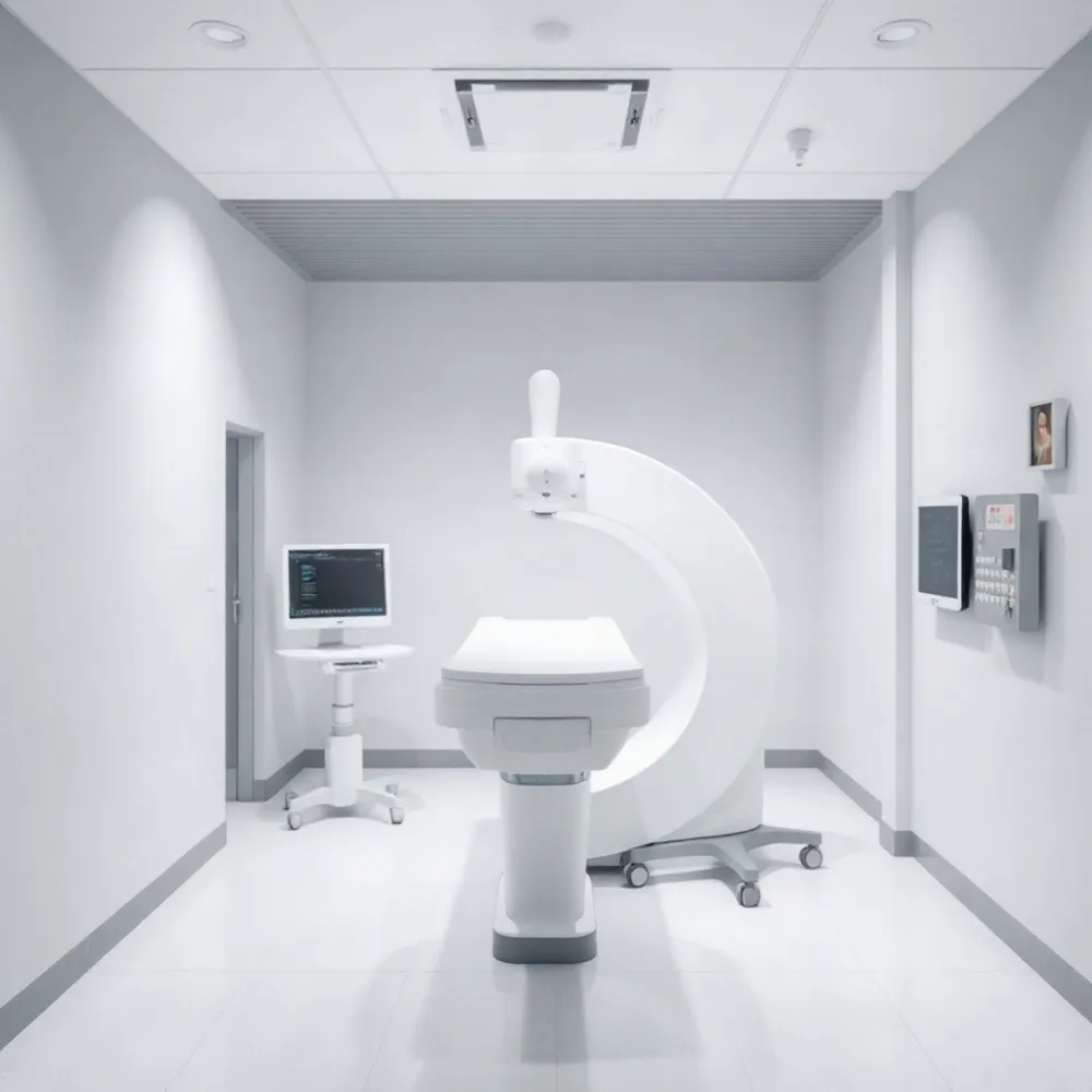 While both MRI and CT scans are essential diagnostic tools for spinal health, they provide different insights based on their underlying technology. CT scans use X-ray technology to create detailed, multi-plane 3D images, making them superior for visualizing complex bony structures, fractures, and severe spinal deformities. In contrast, MRI scans utilize magnetic fields and radio waves to generate comprehensive views of soft tissues, such as spinal discs, nerves, ligaments, and the spinal cord. Because MRI is highly effective at identifying tissue abnormalities without the use of radiation, it is the gold standard for diagnosing conditions like herniated discs or nerve impingement. Ultimately, at osistl.com, your physician may recommend one or both tests to gain a complete picture of your spine structure and surrounding internal anatomy.
How do MRI and CT scans differ in their ability to image the spine?
Selecting the right modality often depends on the suspected injury type. CT scans are frequently preferred for assessing bony alignment and complex fractures due to their high detail for hard tissue, though they involve exposure to ionizing radiation. Conversely, MRI does not use radiation, making it the preferred method for viewing the spinal cord itself and checking for soft tissue injury. If a standard imaging study proves inconclusive, specialists may suggest a myelogram to better highlight concerns within the spinal canal.
What can patients expect during a spinal MRI scan?
During a spinal MRI, you will lie on a motorized table that slides into the scanner magnetic bore, where you must remain very still to ensure clear imaging. While the procedure is painless, you may hear loud tapping, thumping, or snapping noises, for which you will be provided with earplugs or headphones. Although you will be alone in the scanning room, a technologist will monitor you through a window and remain in constant communication via an intercom. The scan typically lasts between 30 to 60 minutes, though it can take longer depending on the specific imaging required. If you feel nervous about the enclosed space or require contrast dye for better image clarity, please discuss these needs with your doctor beforehand, as accommodations can often be made to keep you comfortable.
- Disclose all medical history, especially implanted devices like pacemakers or cochlear implants, as strong magnetic fields pose risks for certain metal items.
- Remove all metal objects, such as jewelry, watches, and eyeglasses, before entering the imaging room.
- Notify your provider if you have a history of contrast allergy or kidney issues, as some scans utilize gadolinium to improve visualization of inflammation or tumors.
- Confirm whether fasting is required, particularly if the procedure involves contrast dye for a CT or MRI study.
Clinical Indicators for Advanced Imaging
 Most spinal discomfort resolves through conservative management, such as physical therapy, activity modification, and targeted exercise. The Orthopedic Spine Institute of St. Louis emphasizes this non-surgical approach as a primary step for patients suffering from back, neck, or sciatica pain. By focusing on conservative recovery initially, specialists can determine if structural healing occurs without the need for invasive procedures.
When is an MRI typically recommended for back pain?
An MRI is typically recommended when a physical examination reveals new or worsening neurological deficits, such as significant muscle weakness, changes in reflexes, or loss of sensation in a specific nerve distribution. It is also essential if you experience sudden bowel or bladder dysfunction, gait abnormalities, or symptoms suggestive of Cauda Equina Syndrome. Furthermore, an MRI is indicated if you have completed at least six weeks of conservative treatment without experiencing meaningful improvement or if your symptoms progress during that time. Additionally, imaging is necessary to rule out or investigate serious underlying conditions, including severe trauma, potential infections, or tumors.
If you are experiencing persistent pain or neurological symptoms, Dr. David S. Raskas and our team at the Orthopedic Spine Institute of St. Louis can evaluate your condition to determine if an MRI is the right next step for your care. While competitors may order advanced imaging immediately, we utilize diagnostic imaging to confirm the precise diagnosis before considering minimally invasive surgical options to ensure that treatment remains as focused and effective as possible.
Neurological Impact of Lumbar and Cervical Spine Injuries
What are the common clinical consequences of damage to the L3, L4, and L5 lumbar vertebrae?
Damage to the L3, L4, and L5 lumbar vertebrae can significantly impact motor and sensory functions in the lower extremities due to their role in nerve innervation and weight support. Patients often experience localized lower back pain, along with numbness, tingling, or radiating discomfort extending into the thighs, legs, and feet. Motor consequences frequently include muscle weakness in the hip flexors, knees, and ankles, which may lead to difficulties with walking, stability, or drop foot. Because these segments house critical nerves, structural issues like herniated discs or stenosis can also lead to impaired reflexes and, in severe cases, disruption of bladder or bowel control. If you are experiencing these symptoms, our team at the Orthopedic Spine Institute of St. Louis is dedicated to providing a thorough evaluation and a patient-first, conservative treatment plan to help restore your quality of life.
What is Brown-Sequard syndrome and how is it characterized?
Brown-Sequard syndrome is a rare, incomplete spinal cord injury caused by damage to one side of the spinal cord, often resulting from trauma such as penetrating wounds, inflammation, or compression. It is clinically characterized by ipsilateral (same-side) motor paralysis, loss of proprioception, and loss of vibration sensation due to damage to the corticospinal tract and posterior columns. Conversely, patients typically experience contralateral (opposite-side) loss of pain and temperature sensation because the spinothalamic tracts decussate, or cross over, shortly after entering the spinal cord. While a pure hemisection of the cord is uncommon, many patients present with a Brown-Sequard-plus syndrome, which involves varying fragments of these sensory and motor deficits. As an incomplete injury, the severity of the neurological deficit can range from mild to severe, often requiring comprehensive multidisciplinary rehabilitation.
At which level of spinal cord injury is there a significant risk for diaphragm failure?
A significant risk for diaphragm failure arises from injuries to the cervical spine at or above the C3 level. This occurs because the phrenic nerves, which are the sole providers of motor function to the diaphragm, originate from the nerve roots at the C3, C4, and C5 levels. Damage to the spinal cord at or above C3 disrupts the essential signals required for these nerves to stimulate diaphragmatic contraction, effectively paralyzing the muscle. Consequently, patients with injuries at this level often face an inability to breathe independently and typically require mechanical ventilation. Because the diaphragm is vital for respiration, understanding these anatomical connection points is critical in evaluating spinal trauma and its impact on pulmonary function. Dr. David S. Raskas and the specialists at the Orthopedic Spine Institute of St. Louis utilize advanced diagnostics to assess these neurological impacts in patients presenting with complex trauma.
Collaborating for Your Spinal Health
Effective spinal care requires more than just high-quality diagnostic images. At the Orthopedic Spine Institute of St. Louis, we integrate these findings with your physical examination, medical history, and clinical symptoms to form an accurate diagnosis. This holistic approach ensures that imaging is used as a tool to guide your recovery rather than an isolated data point.
Dr. David S. Raskas leads our patient-centered team in prioritizing a conservative-first treatment path. By using imaging to confirm structural concerns, we can determine whether physical therapy or activity modification is appropriate or if minimally invasive surgery is necessary to address your specific condition. You can access our centralized digital portal to review our available services, check insurance information, or complete your intake forms to schedule a consultation.